Star News


Baby skeleton. The skeletal system of a child
When a pediatrician examines a newborn in the maternity hospital, he treats him very carefully and carefully and, among other indicators, checks if the baby has any pathologies in the development of joints and bones.
Features of the structure of the bone tissue of a newborn baby
The skeleton of a newborn consists of 50% cartilaginous elements that provide the baby's ability to grow. With age, cartilage is gradually transformed into bone, and this process, as a rule, continues until the age of 18, and its complete completion should occur only by the age of 23-25.The bone tissue of a newborn baby is contained exclusively in the tubular bones, the remaining elements of its skeleton contain only tiny ossification points, which will increase as it grows.
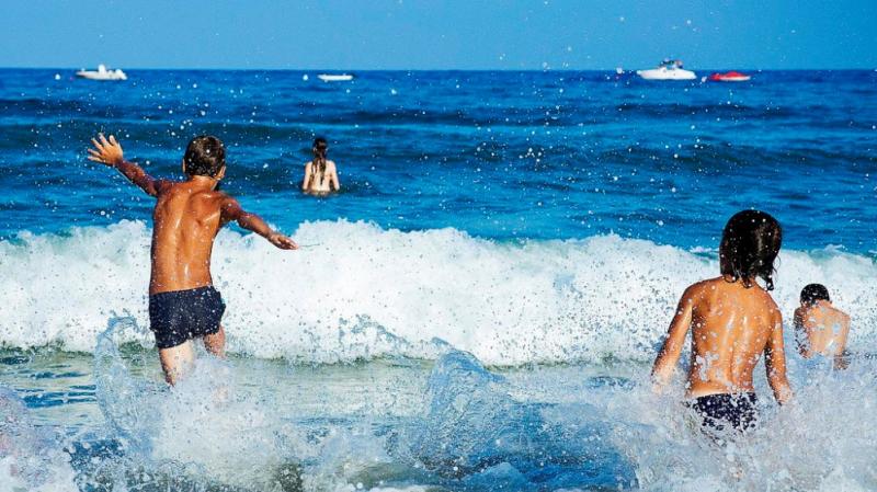
Such a structure of the infant's skeletal system makes it hyperplastic, thanks to which he was able to pass through the mother's birth canal. At the same time, the skeleton of a newborn is vulnerable to such an extent that it can be deformed even with prolonged exposure to gravitational forces. For this reason, experts recommend changing the position of the baby from time to time and not carrying him in the same position in his arms. Newborn babies must be periodically shifted to different hands and turned from side to side. It is not recommended to put the baby on his feet too early, wait until he is physically mature for this. This also applies to the early planting of the child in the pillows. These experiments usually lead to deformation of the baby's skeleton or individual bones.
How does a child's skeleton grow?
The bone tissue of a newborn baby is mainly a coarse-fibrous bundle system, in the mass of which bone plates are randomly located in a small number. Unlike an adult, whose bones have cavities filled with yellow brain, in infants these cavities are tiny and filled mainly with red bone marrow, through which the child's skeleton is supplied with substances necessary for further growth.The epiphyseal cartilage ensures the growth of the child's bones in length. The peripheral edge of this cartilage remains in an active state until almost twenty-five years of age, thanks to which the bones of a person have the opportunity to grow in length and people become taller. But the periosteum is responsible for the growth of bones in width and their thickening. In babies, it is thick, dense and has great functional activity.
For a child, this feature of the periosteum has very favorable moments, even if, God forbid, the baby has a fracture, this tissue remains intact, and the bone protected by it fuses very quickly and without pathological consequences for the child's musculoskeletal system.
Related Articles |
| Victoria Nikitina 20.06 15:04 |
|
I would rather call the bones and joints of a newborn not fragile, but soft, plastic and even flexible. It is especially important to control the correct formation of the hip joints. Therefore, putting the little one on his tummy, taking him by the shins, try to bend his legs at the knees and spread them apart. His posture should resemble that of a frog. The hips should be located almost parallel to the surface of the table. And the ass should go down, and not ride up, like a chicken. Dimples located symmetrically should be visible on the lower back. If you cannot easily perform this exercise, then you should immediately consult an orthopedist, take an x-ray of the hip joints and, possibly, put on stirrups. |
The skeletal system of a baby is very vulnerable. When examining a newly born baby in the maternity hospital, the doctor treats the child very carefully, among other indicators, checking for congenital pathologies in the development of joints and bones.
Features of the structure of the joints of the newborn
In a newborn, the joints are very similar in structure to the joints of an adult, but the skeletal system is quite different. Only about 50% of the constituent bones can be attributed to ash substances only. Everything else is mainly cartilaginous elements that enable the growth of the baby and gradually decrease in volume. This process usually lasts until the age of 18, and is finally completed only by the age of 25.
The basis of the articular and bone tissue of the newborn consists of cartilage. The mobility of the elements forming the joints also has differences. Since the joints have not yet been developed in a newborn, the range of motion is very small, while the probability of dislocations is quite high in case of careless handling. This immaturity of the joints persists, as a rule, up to three to five years, until the bone and joint tissue develops sufficiently, and the child does not fully learn to control his body.

In a newborn, the structure of bone tissue also has certain differences. The bones of a newborn are a coarse fibrous bundle system. While in the bones of an adult there are large cavities that are filled with yellow marrow, in an infant these cavities are quite small and mainly contain red marrow. It is thanks to a significant amount of this brain that the baby’s skeletal system is supplied with blood enough for growth. This process is very intense until about two years of age. After a certain decline, with renewed vigor, the growth process resumes already in the puberty period.
In length, bone growth is provided by the epiphyseal cartilage, the peripheral edge of which remains active right up to the age of twenty-five, thanks to which the bones can increase in length, and the child becomes taller.
The periosteum is responsible for the growth of bones in width. In a baby, it is thick, dense and more functionally active. This feature of the periosteum is quite favorable for children, since it is often not damaged during fractures, and the bone protected by it fuses faster and without consequences for the child's musculoskeletal system.
In a newborn, bone tissue is contained only in tubular bones, while other elements of the skeleton have only small areas of ossification, which increase as the child grows.
The skeleton is the skeleton of the body, consisting of 206 bones, which act as levers for the muscles and create counter-traction for them, thereby providing movement. The skeleton is a rigid structure that supports and protects the body by surrounding and enclosing the vital organs in the head, chest, and abdomen. The skeleton of a newborn consists mainly of cartilage - soft, fibrous and elastic tissue, from which, together with the bones, a mature skeleton is formed.
The hard outer part of the bone consists of cells arranged in thousands of cylinders, which help to evenly distribute the stings that act on the bone. In the bone marrow, located in the middle of the bone, most of the leukocytes (white blood cells) and all the erythrocytes (red blood cells) are formed. In young children, all bones contain hematopoietic bone marrow (in adults, bone marrow functions only in the bones of the body). Bones contain salts, mainly calcium and phosphorus, which contribute to their strength and rigidity.
How bones develop
In childhood, at certain stages of development, including adolescence, cartilage tissue turns into bone, and then the skeleton is finally formed.
Bone is a living but hard tissue that grows, develops and renews itself. Old bone cells are constantly resorbed and new ones are formed in their place. In childhood, there is a continuous reconstruction of the skeleton and its strengthening. Tubular bones grow mainly in the region of the epiphysis, or in the "growth zone" of the bone. Injury to this area can impair growth. The development of healthy bones depends both on exercise and on sufficient dietary intake of vitamins (especially vitamin D), salts (primarily calcium), and protein.
X-rays do not show what happens to the bones as they form. At birth, each bone has its own outline. The bones of a child's hand are made up of cartilage (not visible on x-ray) and bone (in the form of dense areas) tissues. As a child develops, cartilage turns into bone. In adolescence, this transformation is completed.
Signs of the disease
In childhood, bone fractures, joint dislocations and muscle strains are most common. Children are more likely to have minor injuries; inflammation and bone tumors are rare. Sometimes there is a curvature of the spine, called scoliosis, for which early diagnosis is necessary for successful treatment. If a child lies still or refuses to move a bruised limb, he is most likely seriously injured: if a child continues to use a bruised hand or arm during play, the damage is unlikely to be serious.
muscles
All movements of the body and internal organs are carried out with the help of muscles. Muscles are made up of thousands of individual fibers that contract to cause movement. There are two types of muscles: voluntary, performing movements of the body itself, and involuntary, producing movements of internal organs (for example, the digestive tract, whose muscles contract rhythmically to move food). Muscles "bloom" from work - physical exercises improve their blood circulation, increase mass and increase efficiency.
A child is born with certain instinctive reactions, which are called reflex movements. Gradually, as the central nervous system and muscles develop, they disappear, which allows the baby to control his body more and more confidently.
How muscles grow
While still in the womb, the baby moves vigorously and continues to do so after birth, using all muscle groups. However, at this stage, the muscles are still underdeveloped and require nutrients, exercise and hormones for their full maturation. In adolescence, male hormones contribute to an increase in the strength and size of the muscles of boys. Physical exercise is essential for proper muscle development; without them, the muscles practically wither. In physically active children, the muscles become stronger and well coordinated.
Signs of the disease
Undertrained or overtrained muscles are more susceptible to damage. With insufficient exercise, the muscles dry out and become flabby. Further inactivity leads to muscle atrophy and weakness. Excessive exercise usually causes pain, stiffness, and sometimes inflammation and swelling of the muscles. Weakness and pain in the muscles can also occur due to a viral infection.
Read other articles in the section
Children are interested in how the world works and everything in it. Their curiosity makes no exception for humans. They are interested in how a person works, how he sees and hears, runs and jumps. About the human skeleton, which you can’t see, like skin or eyes, with the naked eye, modern children learn from cartoons and comics. This makes the skeleton in the eyes of the child even more interesting.
But the human skeleton with the name of the bones and muscles in cartoons and comics can not be found, and it will not hurt children to memorize them little by little.
Knowing how complex and fascinating the human body is, will arouse in the child an interest in biology, medicine, and will encourage a more conscious approach to their own health and the health of others. Finally, this knowledge will be useful to him at school, where already in the elementary grades they get acquainted with the structure of man.
The skeleton and muscles are the frame that determines the shape of a person, protects his internal organs and allows him to move. If not for the skeleton, then the person would be like a shapeless jellyfish. Muscles are attached to the skeleton and provide our every movement - from fluttering eyelashes to lifting weights.

Bones are composed of organic and inorganic substances, the former of which provide them with flexibility, and the latter with strength. Thanks to this, the bones are unusually elastic and strong. The complex structure adds strength and flexibility to them at the same time. Any bone consists of several layers.
- The outer one is made up of strong bone tissue.
- The next connecting layer covering the outside of the bone.
- Loose connective tissue containing blood vessels.
- At the ends is cartilage tissue, due to which bone growth occurs.
- Another layer is the nerve endings, through which signals are transmitted from the brain and back.
Inside the bone tube is the bone marrow, which also comes in two forms. Red is involved in hematopoiesis and bone formation. It is full of blood vessels and nerves. Yellow is responsible for the growth and strength of bones. We see that the skeleton, in addition to everything else, contributes to the renewal of blood. This is where blood cells are born. If, due to illness, it ceases to perform this task, the organism dies.
In the organization of the skeleton, several groups of bones are distinguished. One of them is the main supporting structure of our body, which includes the spine, bones of the head and neck, chest and ribs. Together they form the axial skeleton. The second part is called the accessory skeleton and it includes the bones that form our arms and legs, and the groups of bones that provide their connection with the axial skeleton.
Skeleton structure
 The bones of the head include the skull and bones of the middle ear. The skull contains and protects the brain. It consists of two sections: brain and facial. The first of which includes eight bones. There are fifteen of them in the front section.
Trunk bones

This part of the skeleton includes the chest and spine, starting from the neck. We unite them, since they are closely related both literally (the chest is attached to the spine), and by location, and by the tasks they solve. These are one of the largest human bones. Their function is to protect the heart, lungs, etc. Among them are the spinal column and chest.
vertebral column

The human spine is the main support of the whole body, its main axis. It is he who ensures our upright posture. The spinal cord provides communication between the upper and lower parts of the body. It distinguishes five departments, consisting of 32-34 vertebrae. They are called by their location - cervical, thoracic, lumbar, sacral and coccygeal.
Rib cage
 The chest really looks like a cage, where 12 pairs of ribs play the role of a lattice behind which the heart, lungs, and vital organs are hidden. Closes its flat wide bone, which is called the sternum. In total, 37 bones belong to the chest.
Upper limb bones

So scientists and doctors call our hands. I don’t think it’s necessary to explain how much it means for a person to be able to perform both weight lifting and cross-stitching. But think about how different tasks they are called upon to solve. This explains their rather complex structure. The bones of the upper limb (VC) include the belt of the VC and the free part of the VC.
The belt includes the scapula and collarbone, connected by a ball joint to the humerus. This is where the muscles come in. In the free part of the upper limb, three sections are distinguished - the shoulder (humerus), forearm (radius and ulna) and the hand. Most of the bones in this particular area of the hand - twenty-seven, they are noticeably smaller than the bones of the forearm, and differ from them in shape.
Pelvic girdle

This belt provides a connection between the spine and lower extremities, and also accommodates and protects the organs of the digestive, urinary and reproductive systems. The pelvis is made up of three fused bones.
Bones of the lower limb
 The skeleton of the leg resembles the structure of the hand. They are fundamentally the same, differing in size and some other details. Since it is the legs that bear the main weight of our body when moving, they are more powerful and stronger than the bones of the arm.
What are the shapes of bones
Depending on their functions in the human body, bones differ in shape. There are four types of bone shapes:
- Wide or flat (for example, at the skull);
- Long or tubular (mainly in the limbs);
- Short, such as the bones of the wrist;
- Asymmetrical, having a composite shape. These are the pelvic bones, vertebrae, etc.
Muscles of the head and face

Previously, only specialists could know the structure of a person, his skeleton and a list of muscles. Today, anyone who is interested in this topic can find a detailed anatomical atlas on the Internet, which provides a detailed description of the movements of our body and all its parts that provide this. The most important role in ensuring movements is played by muscles, organs consisting of a special elastic tissue capable of
contract under the influence of nerve impulses. There are over 640 different muscles in the human body. Among them, there are various types according to different parameters:
- By the functions they provide;
- In the direction of the fibers of which they are composed;
- By form;
- In relation to the joints.
Understanding all this is not so easy, so let's look at the muscles depending on where they are on our body.
When we talk about movement, we first of all imagine how our arms and legs work. Meanwhile, the muscles of the head and face also work hard, providing breathing, facial expressions, speech, and our nutrition. The strongest muscles in our body are the chewing muscles.
The facial muscles and muscles of the eye, unlike all the others, are not attached to the bones. This allows them to be particularly sensitive and guarantee even micro-movements. Thanks to this, we can convey both joy and sadness, the slightest change in emotions.
Neck muscles
 This group of muscles allows us to turn around, bow, swallow something and speak, even breathe.
Trunk muscles

Muscles are attached to bones by tendons and perform various tasks. - provide mobility and the ability to maintain balance, fix the joints. According to their functions and modes of action, there are those that contract synchronously during work or synergists, and muscles that perform opposite actions (antagonists). Most often, actions occur due to the fact that some muscles contract and other muscles relax at the same time.
The muscles of the trunk include the superficial and deep muscles of the back and chest, oblique, rectus, etc. abdominal muscles.
Pelvic muscles

These muscles originate on the bones of the pelvis and spine, are attached to the upper edge of the thigh, and surround the hip joint. Among them, two groups are distinguished: internal and external.
Muscles of the upper limbs

Among this group of muscles, the same parts are distinguished as in the bones of the hand:
- Muscles of the VK belt;
- shoulder;
- Forearms, providing flexion and extension of the forearm, hand and each finger.
Muscles of the lower limbs

Thanks to these muscles, a person walks and runs, swims or jumps. In order to provide such different actions, no single group of different muscles is required. These include the muscles of the thigh, lower leg and foot. This is a rather complex system, including muscles that are different in shape, the direction of the fibers, in relation to the joints and other things, mutually complementing each other.
Muscle Anatomy Muscle Physiology How Muscles Work
The skeleton is the backbone of the whole organism. Separate parts of the skeleton serve to protect such important organs as the brain, heart, lungs, etc. In addition, the skeletal system, in combination with the muscular system, forms the organs of human movement, while the bones are levers actuated through the muscles attached to them. The nervous system gives impulses to muscle contraction.
The skeleton of a child is laid down in the early uterine period and consists mainly of cartilage tissue. Even in the uterine period, cartilage tissue begins to be replaced by bone tissue. The process of ossification proceeds gradually, and not all bones of the skeleton ossify at the same time. The ossification process is completed by the age of 20-25.
Changes occur in the chemical composition of bone tissue throughout a person's life up to a very old age. At younger ages, there are very few calcium and phosphorus salts in the bone tissue. Due to the fact that there are few calcium salts in the bones of children, and organic elements predominate, and the processes of ossification are far from complete, the skeleton of children has great elasticity and can easily be subjected to curvature.
The spine in an adult has three curvatures. One of them - the cervical - has a bulge forward, the second - the thoracic - is bulging back, the third - the lumbar curvature is directed forward. In a newborn, the spinal column has almost no bends. The first cervical curvature is formed in a child already when he begins to hold his head on his own. The second in order is the lumbar curvature, which also faces forward with a bulge when the child begins to stand and walk. The thoracic curvature, bulging backwards, is the last to form, and by the age of 3-4 years, the child's spine acquires curves characteristic of an adult, but they are not yet stable. Due to the great elasticity of the spine, these curves are smoothed out in children in the supine position. Only gradually, with age, the curvature of the spine becomes stronger, and by the age of 7, the constancy of the cervical and thoracic curvature is established, and by the onset of puberty, the lumbar curvature.
Only gradually, as the child grows, does the process of ossification of the spine occur. Until the age of 14, the spaces between the vertebral bodies are still filled with cartilage. At the age of 14-15, new ossification points appear between the vertebrae in the form of thin plates on the upper and lower surfaces of the vertebrae. Only by the age of 20 do these plates fuse with the vertebral body. The line of their fusion remains pronounced until the age of 21. The tops of the transverse and spinous processes of the vertebrae up to 16-20 years also remain covered with cartilage when ossification points appear on them. Fusion of cartilaginous plates with arches is completed after 20 years.
These features of the development of the spine of a child and adolescent cause its slight compliance and possible curvature in case of incorrect body positions and prolonged stress, especially unilateral. In particular, curvature of the spine occurs when sitting incorrectly on a chair or at a desk, especially in cases where the school desk is improperly arranged and does not correspond to the height of children; when sleeping for a long time with a bent torso on one side, etc. Curvature of the spine can be in the form of a bend in the cervical (especially in infants if they are not carried properly on their hands) and thoracic parts of the spine to the side (scoliosis). Scoliosis of the thoracic spine most often occurs at school age as a consequence of improper seating. Antero-posterior curvature of the thoracic spine (kyphosis) is also observed as a result of prolonged improper seating. Curvature of the spine can also be in the form of excessive curvature in the lumbar region (lordosis). That is why school hygiene attaches so much importance to a properly arranged desk and imposes strict requirements on the seating of children and adolescents.
Fusion of segments of the sternum also occurs relatively late. So the lower segments of the sternum grow together at the age of 15-16, and the upper segments only by the age of 21-25, and only the handle of the sternum remains independent. In case of prolonged improper landing, in cases where a child or teenager rests his chest on the edge of the desk cover, a change in the chest may occur and disturbances in its development may occur. This, in turn, adversely affects the normal development and activity of the lungs, heart and large blood vessels located in the chest.
The development of the pelvic bones in children, especially girls, is also of hygienic interest. The adult pelvis consists of two nameless bones and a sacrum wedged between them. The latter represents five pelvic vertebrae fused together. The pelvis in children is different in that each innominate bone consists of three independent parts adjacent to each other: the ilium, ischium and pubis. Only from about the age of 7 do these bones begin to fuse with each other, and the process of their fusion basically ends by the age of 20-21, when the nameless bone becomes one. This circumstance must be taken into account, especially in relation to girls, since their genitals are enclosed in the pelvis. With sharp jumps from a great height onto a hard surface, an imperceptible displacement of the pelvic bones that have not yet fused and, subsequently, their incorrect fusion can occur.
The wearing of high-heeled shoes by adolescent girls also contributes to the change in the shape of the pelvis. The human foot has the shape of an arch, the bases of which are the back stop of the calcaneus, and in front - the heads of the first and second metatarsal bones. The arch has the ability of elastic stretching, "springs", due to which impacts on the soil are softened. Narrow shoes, tightening the foot, make it difficult for the arch to work like a spring and lead to the formation of a flat foot (the arch is smoothed out). High heels change the shape of the arch and the distribution of the load on the foot, shift the center of gravity forward, as a result of which you have to tilt your torso back so as not to fall forward when walking. The constant wearing of shoes with high heels leads to a change in the shape of the pelvis. With incompletely fused pelvic bones, this deviation of the body and a shift in the center of gravity can lead to a change in the shape of the pelvis, and, moreover, in the direction of reducing the outlet of the pelvic cavity due to the approach of the pubic bones to the sacrum. It is quite obvious that for a girl, when she becomes a woman, this curvature of the pelvis can become fatal and adversely affect the birth function.
The cranial bones of the newborn are also in the stage of ossification and have not yet fused together, with the exception of the upper jaw and premaxilla. The cranial bones are connected to each other by a soft connective tissue membrane. Between them there are places that are not yet covered with bone tissue, peculiar membranous spaces - large and small fontanels, covered with connective tissue. A small fontanel overgrows by 2-3 months, and a large one by 1 year is already covered with bone tissue. The cranial sutures finally fuse only by 3-4 years, sometimes later. In children at an early age, the brain part of the skull is more developed than the facial part.
The bones of the skull grow most intensively during the first year. In subsequent years, the growth of the skull occurs unevenly: periods of strong growth are replaced by periods of relative calm. Thus, a relatively strong growth of the skull occurs from birth to 4 years, from 6 to 8 years and from 11 to 13 years. From 7 to 9 years there is a strong growth of the base of the skull. In the period from 6 to 8 years, a strong development of the facial part of the skull is already noticeable. But the most intensive development of the facial part of the skull begins from 13 to 14 years of age and then proceeds during puberty, when the final relationship between the brain and the facial part of the skull is established.
The ossification of the tubular bones that make up the skeleton of the limbs begins in the fetal period and proceeds extremely slowly. Inside the middle part of the tubular bone (diaphysis), a cavity is formed, which is filled with bone marrow. The ends of long tubular bones (epiphyses) have their own separate ossification points. Complete fusion of the diaphysis and epiphyses is completed at the age of 15 to 25 years.
The development of the process of ossification of the hand is of great importance in hygienic terms, since through the hand the child learns to write and perform various labor movements. The newborn has no carpal bones at all yet and they are only just emerging. The process of their development proceeds gradually, and they become clearly visible, but not yet fully developed, only in children of 7 years old. Only by the age of 10-13 the process of ossification of the wrist is completed. The process of ossification of the phalanges of the fingers ends by 9-11 years.
These features of ossification of the hand are important for the correct organization of teaching children to write and work. It is quite obvious that for a child's not completely ossified hand, it is necessary to give a pen that is accessible to him in size and shape for writing. In this regard, it becomes clear that quick (fluent) writing is not possible for children of elementary grades, while for adolescents, in whom the process of ossification of the hand ends, as a result of a gradual and systematic exercise, fluent writing becomes available.
It can be seen from the foregoing that not only in children of younger ages, but also in adolescents in high school, the processes of ossification are not yet fully completed and in many parts of the skeleton they continue until the period of adulthood. The described features of bone development in children and adolescents put forward a number of hygienic requirements, which have already been partially indicated above. Due to the fact that the process of ossification of the skeleton of a child of preschool and school age has not yet been completed, improper organization of educational work and forcing the child to exercises of the motor apparatus beyond his age can bring him great harm and cause mutilation of the child's skeleton. Especially dangerous in this regard are excessive and one-sided physical stresses.
Moderate and affordable physical exercises for children, on the contrary, are one of the means of strengthening bone tissue. Extremely important for a growing body are physical exercises associated with respiratory movements and entailing the expansion and collapse of the chest, since they contribute to its growth and strengthening of bone tissue.
Exercises of the upper and lower extremities intensify the processes of growth of long bones, and, conversely, lack of movement, pressure on the bone tissue (by swaddling, clothes squeezing the body, etc.), incorrect body position entail a slowdown in the growth of bone tissue. The development of bones, their chemical composition and strength are influenced by the nutritional conditions and the environment surrounding the child and adolescent.
For the normal development of bone tissue in children, the presence of good-quality air, an abundance of light (especially constant access to direct sunlight), free movements of all members of the body and rational nutrition of the body are necessary.