Star news


Placenta previa - classification, symptoms, diagnosis, principles of treatment. Childbirth with placenta previa
But the diagnosis of “placenta previa” is not a reason to panic - it only means that the expectant mother needs to take care of herself and not neglect the doctor’s recommendations.
During the normal course of pregnancy, the placenta (the organ that supplies blood, and with it oxygen and nutrients to the fetus) is usually located in the fundus (upper part of the uterus) or on the walls of the uterus, usually along the back wall, with transition to the side walls, those. in those areas where the walls of the uterus are best supplied with blood. On the anterior wall, the placenta is located somewhat less frequently, since the anterior wall of the uterus undergoes much greater changes than the posterior one. In addition, the location of the placenta along the posterior wall protects it from accidental injury.
Placenta previa is a pathology in which the placenta is located in the lower parts of the uterus along any wall, partially or completely covering the area of the internal os - the area of the exit from the uterus. If the placenta only partially covers the area of the internal os, then this is an incomplete presentation, which is noted with a frequency of 70-80% of the total number of presentations. If the placenta completely covers the area of the internal os, then this is called complete placenta previa. This option occurs with a frequency of 20-30%.
There is also a low location of the placenta, when its edge is at a lower level than it should be normally, but does not overlap the area of the internal os.
Causes
The most common reasons for the formation of a low-lying placenta or placenta previa are pathological changes in the inner layer of the uterus (endometrium) due to inflammation, surgical interventions (curettage, cesarean section, removal of myomatous nodes - nodes of a benign tumor of the uterus, etc.), multiple complicated births. In addition, disturbances in placental attachment may be caused by:
- existing uterine fibroids;
- endometriosis (a disease in which the inner lining of the uterus - the endometrium - grows in uncharacteristic places, for example, in the muscle layer);
- underdevelopment of the uterus;
- isthmic-cervical insufficiency (a condition in which the cervix does not perform its obturator function, it opens slightly and the fertilized egg is not retained);
- inflammation of the cervix;
- multiple pregnancy.
Due to these factors, the fertilized egg entering the uterine cavity, after fertilization, cannot be implanted in a timely manner in the upper parts of the uterus, and this process occurs only when the fertilized egg has already descended into its lower parts. It should be noted that placenta previa is more typical for repeatedly pregnant women than for first-time mothers.
How does placenta previa manifest?
The most common manifestation of placenta previa is repeated bleeding from the genital tract. Bleeding can occur during various periods of pregnancy, starting from the earliest stages. However, most often they are observed in the second half of pregnancy. In the last weeks of pregnancy, when uterine contractions become more intense, bleeding may increase.
The cause of bleeding is the repeated abruption of the placenta, which is not able to stretch following the stretching of the uterine wall as pregnancy progresses or labor begins. In a normal position, the placenta is located in those areas of the uterus that are subject to the least stretching. In this case, the placenta partially detaches, and bleeding occurs from the vessels of the uterus. The fetus does not lose blood. However, he is at risk of oxygen starvation, since the detached part of the placenta does not participate in gas exchange.
Provoking factors for bleeding with placenta previa or low placenta can be: physical activity, sudden coughing, vaginal examination, sexual intercourse, increased intra-abdominal pressure with constipation, thermal procedures (hot bath, sauna).
With complete placenta previa, bleeding often appears suddenly, i.e. without provoking factors, without pain, and can be very abundant. Bleeding may stop, but after some time it may recur, or it may continue in the form of scanty discharge. In the last weeks of pregnancy, bleeding resumes and/or intensifies.
With incomplete placenta previa, bleeding may begin at the very end of pregnancy, but more often it occurs at the beginning of labor. The severity of bleeding depends on the size of the presenting area of the placenta. The more placental tissue is present, the earlier and more severe the bleeding begins.
Repeated bleeding during pregnancy complicated by placenta previa, in most cases, leads to the development of anemia - a decrease in the amount of hemoglobin in the blood.
Pregnancy with placenta previa is often complicated by the threat of miscarriage; this is due to the same reasons as the occurrence of improper location of the placenta. Preterm birth most often occurs in patients with complete placenta previa.
Pregnant women with placenta previa are characterized by low blood pressure, which occurs in 25-34% of cases,
Management of pregnant women in an obstetric hospital involves, if necessary, the use of medications that eliminate contractile activity of the uterus.
Preeclampsia (a complication of pregnancy characterized by disruption of all organs and systems of the expectant mother, deterioration of uteroplacental circulation, often manifested by increased blood pressure, the appearance of protein in the urine, and edema) is also no exception for pregnant women with placenta previa. This complication, occurring against the background of dysfunction of a number of organs and systems, as well as with the phenomena of blood clotting disorders, significantly worsens the nature of repeated bleeding.
Placenta previa is often accompanied by fetoplacental insufficiency (the fetus receives insufficient oxygen and nutrients) and fetal growth retardation. The detached part of the placenta is excluded from the general system of uteroplacental circulation and does not participate in gas exchange. With placenta previa, an abnormal position of the fetus (oblique, transverse) or breech presentation often occurs, which, in turn, is accompanied by certain complications.
What is “placenta migration”
In obstetric practice, the term “placental migration” has become widespread, which, in fact, does not reflect the real essence of what is happening. The location of the placenta changes due to changes in the structure of the lower segment of the uterus during pregnancy and the direction of placental growth towards better blood supply to areas of the uterine wall (towards the fundus of the uterus) compared to its lower sections. A more favorable prognosis in terms of placental migration is noted when it is located on the anterior wall of the uterus. Typically, the process of “placenta migration” occurs within 6 weeks and is completed by 33~34 weeks of pregnancy.
Diagnostics
Detecting placenta previa is not particularly difficult. The presence of placenta previa may be indicated by a pregnant woman's complaints of bleeding. In this case, repeated bleeding in the second half of pregnancy is usually associated with complete placenta previa. Bleeding at the end of pregnancy or at the beginning of labor is most often associated with incomplete placenta previa.
If there is bleeding, the doctor will carefully examine the vaginal walls and cervix using speculum to rule out trauma or pathology of the cervix, which may also be accompanied by bleeding.
A vaginal examination of a pregnant woman also easily reveals clear diagnostic signs indicating an abnormal location of the placenta. Currently, the most objective and safe method for diagnosing placenta previa is ultrasound, which allows you to establish the fact of placenta previa and the variant of presentation (complete, incomplete), determine the size, structure and area of the placenta, assess the degree of abruption, and also obtain an accurate idea of placental migration.
If an ultrasound reveals complete placenta previa, then a vaginal examination is not performed at all, as it can cause bleeding. The criterion for a low location of the placenta in the third trimester of pregnancy (28-40 weeks) is the distance from the edge of the placenta to the area of the internal os of 5 cm or less. Placenta previa is indicated by the detection of placental tissue in the area of the internal os.
The nature of the location of the placenta in the second and third trimesters of pregnancy (up to 27 weeks) is judged by the ratio of the distance from the edge of the placenta to the area of the internal os with the diameter of the fetal head.
If an abnormal location of the placenta is detected, a dynamic study is carried out to monitor its “migration”. For these purposes, at least three times echographic control (ultrasound) is required throughout pregnancy at 16, 24-26 and 34-36 weeks.
Ultrasound should be performed when the bladder is moderately full. Using ultrasound, it is also possible to determine the presence of blood accumulation (hematoma) between the placenta and the wall of the uterus during placental abruption (in the event that there is no bleeding from the uterine cavity). If the area of placental abruption occupies no more than 1/4 of the area of the placenta, then the prognosis for the fetus is relatively favorable. If the hematoma occupies more than 1/3 of the area of the placenta, then most often this leads to the death of the fetus.
Features of pregnancy management and childbirth
The nature of pregnancy in women with placenta previa depends on the severity of bleeding and the amount of blood loss.
If there is no bleeding in the first half of pregnancy, then the pregnant woman can be at home under outpatient monitoring in compliance with a regimen that excludes the action of provoking factors that can cause bleeding (limitation of physical activity, sexual activity, stressful situations, etc.).
Observation and treatment for pregnancy over 24 weeks is carried out only in an obstetric hospital in any case, even in the absence of bleeding and normal health.
Treatment aimed at continuing pregnancy until 37-38 weeks is possible if the bleeding is not heavy and the general condition of the pregnant woman and fetus is satisfactory. Even despite the cessation of bleeding from the genital tract, a pregnant woman with placenta previa cannot under any circumstances be discharged from the hospital before giving birth.
Management of pregnant women in an obstetric hospital includes:
- adherence to strict bed rest;
- if necessary, use medications to eliminate contractile activity of the uterus;
- treatment of anemia (reduced amount of hemoglobin) and fetoplacental insufficiency.
If the pregnancy was carried to 37-38 weeks and placenta previa persists, depending on the current situation, the optimal method of delivery is selected individually.
The absolute indication for elective caesarean section is complete placenta previa. Childbirth through the vaginal birth canal is impossible in this situation, since the placenta blocking the internal os does not allow the presenting part of the fetus (this can be the fetal head or the pelvic end) to be inserted into the entrance to the pelvis. In addition, as uterine contractions increase, the placenta detaches more and more, and bleeding increases significantly.
In case of incomplete placenta previa and in the presence of accompanying complications (breech presentation, abnormal position of the fetus, uterine scar, multiple pregnancy, severe polyhydramnios, narrow pelvis, primigravida over 30 years of age, etc.), a caesarean section is also performed as planned.
If the above-mentioned associated complications are absent and there is no bleeding, then the doctor waits until spontaneous labor begins and opens the amniotic sac. If, after opening the amniotic sac, bleeding still begins, the issue of performing a cesarean section is decided.
If, with incomplete placenta previa, bleeding occurs before the onset of labor, then the amniotic sac is also opened. The necessity and expediency of this procedure is due to the fact that when the membranes are opened, the fetal head is inserted into the entrance to the pelvis and presses the detached part of the placenta against the wall of the uterus and pelvis, which helps to stop further placental abruption and stop bleeding. If bleeding continues after opening of the membranes and/or the cervix is immature, then a cesarean section is performed. If the bleeding stops and there are no complications, delivery through the natural birth canal is possible.
Bleeding can begin in the early stages of labor, from the moment of the first contractions. In this case, the fetal bladder is also opened.
Thus, management of childbirth through the natural birth canal with incomplete placenta previa is possible if:
- bleeding stopped after opening of the amniotic sac;
- the cervix is mature;
- labor activity is good;
- there is a cephalic presentation of the fetus.
However, cesarean section is one of the most frequently chosen methods of delivery by obstetricians for placenta previa and is performed for this pathology with a frequency of 70-80%.
Other typical complications during childbirth with incomplete placenta previa are weakness of labor and insufficient oxygen supply to the fetus (fetal hypoxia). A mandatory condition for conducting childbirth through the natural birth canal is constant monitoring of the condition of the fetus and contractile activity of the uterus; Sensors are attached to the woman’s stomach, which are connected to a device that records the fetal heartbeat and the presence of contractions; these parameters are recorded on tape or projected onto a monitor.
After the birth of the child, bleeding may resume due to a disruption in the process of separation of the placenta, since the placental site is located in the lower parts of the uterus, the contractility of which is reduced.
Heavy bleeding often occurs in the early postpartum period due to decreased uterine tone and damage to the extensive vascular network of the cervix.
Prevention of placenta previa involves the rational use of contraception, the exclusion of abortions, and the early detection and treatment of various inflammatory diseases of the reproductive system and hormonal disorders.
As doctors said, the placenta is provided by nature specifically for the growth and development of the fetus. It’s not for nothing that its name translated from Latin means “children’s place.” For the normal course of pregnancy, not only the structure, but also the location of this organ is important.
However, the diagnosis of “marginal placenta previa” is not yet a reason to become despondent. It only means that the expectant mother needs to be more attentive to her health.
How the placenta is formed
Briefly, the mechanism of the appearance of the placenta can be described as follows. Once in the uterine cavity, the fertilized egg is immersed in its mucous membranes like a ball in thick sour cream. By the ninth day, villi form on the surface of the embryo, which grow into the wall of the uterus. Subsequently, the placenta begins to develop from them. The final formation of the organ occurs by 13-16 weeks of pregnancy.
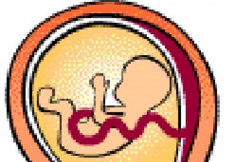
The shape of the placenta is similar to a flat disc, to the center of which the umbilical cord is attached. Incredibly important processes for the baby take place inside this disk. Here it receives nutrients and oxygen from the mother. This is also where the toxins “used up” by the child’s body are removed: carbon dioxide and other waste. In essence, the placenta serves as lungs, intestines and kidneys in “one bottle” for the fetus.
What is marginal presentation
Normally, the placenta forms in the upper part (at the “bottom”) of the uterus along its posterior, lateral, or, less commonly, anterior wall. This position protects her from accidental injury.
The presentation of an organ (placenta praevia) is said to occur when it is formed in the lower part of the uterus next to the outlet - the pharynx. Literally, this term is translated from Latin as “on the path before life,” that is, the placenta becomes an obstacle to the birth of the baby.
There are three types of presentation:
- complete (the placenta covers the pharynx 100%),
- lateral (the hole is blocked by 50-70%),
- edge (the exit is closed by no more than 30%).
Of all the options, marginal presentation is considered the most favorable, since in this case there is a high probability that the birth will take place naturally. Pathology is usually detected during a routine ultrasound.
Who is at risk?
In most cases, abnormal position of the placenta is associated with pathological changes in the inner layer of the uterus. Because of this, the fertilized egg cannot “catch” onto the upper part of the endometrium and goes down.
The causes of such conditions may be:
- abortion,
- surgical interventions that preceded the current pregnancy (curettage of the uterine cavity, cesarean section, removal of benign nodes),
- sexually transmitted infections
- inflammation of the cervix.
The development of pathology can also be caused by congenital anomalies of the uterus or fibroids, which caused its deformation.
In multiparous women, marginal presentation is diagnosed three times more often than in women who are about to give birth to their first child. Perhaps this is due to the fact that experienced mothers manage to suffer a greater number of gynecological diseases by the time of their second or third birth.
Risks of complications
Why is marginal placenta previa dangerous? Nothing in the early stages. The baby is growing and developing normally. Mom doesn’t feel her pathology at all.
Problems may arise in the third trimester, when the uterus greatly increases in size. The tissue of the placenta has little elasticity, so it does not have time to stretch after the endometrium. In some places, the organ detaches from the walls of the uterus, which leads to damage to blood vessels. Blood flows out through the cervical canal.
The fetus does not suffer. However, with excessively frequent bleeding, it begins to receive less oxygen, since the detached areas of the placenta cease to function.
Bleeding can be caused by:
- physical exercise,
- sexual contact,
- hot procedures (bath, sauna),
- examination by a gynecologist,
- straining of the abdomen.
With marginal presentation, the discharge is light and painless. Sometimes they appear at night for no reason.
Bleeding may begin in the last weeks of pregnancy. But in most cases, they do not bother the woman until the onset of labor, when the contractile activity of the uterus sharply increases.
It happens that marginal presentation is accompanied by an incorrect position of the fetus - oblique, pelvic or transverse. In this case, the birth process may become more complicated.
Observation and treatment
In the early stages, patients are observed on an outpatient basis. No drug therapy is provided. A woman is recommended:
- avoid stress, physical activity, sexual intercourse,
- maintain a healthy diet,
- have a full rest.
It should be noted that the final diagnosis occurs only closer to childbirth. Until this time, there remains a possibility that the position of the placenta will change to normal. This is all connected with the same increase in the size of the uterus. Stretching, the endometrium pulls the placenta along with it, and its edges move away from the pharynx area.
If bleeding occurs, the patient is hospitalized and further observation is carried out in a hospital setting.
How the birth will occur with marginal placenta previa is decided by the doctor in the last weeks of gestation. If there are associated complications (polyhydramnios, narrow pelvis, kidney disease, uterine scar), a cesarean section is performed. When women feel well, they tend to opt for natural childbirth.
If a pregnant woman experiences bleeding before labor begins, the amniotic sac is opened. This prevents further placental abruption and helps stop the discharge. If the bleeding cannot be stopped, the birth is completed surgically.
A mandatory condition for childbirth is continuous monitoring of the condition of the woman and baby. Sensors are attached to the patient's abdomen that record the fetal heartbeat.
After the baby is born, severe bleeding is also possible. In this case, doctors have to resort to surgical removal of the placenta.
(3 Votes)
The placenta begins its development starting in the first week of pregnancy and is fully formed at the beginning of the second trimester. Thanks to it, the fetus is nourished and receives oxygen, metabolism occurs and, generally speaking, the placenta ensures a successful pregnancy. Therefore, this organ is very important for a pregnant woman and any deviations in the development of the placenta must be monitored in a timely manner.
One of the deviations is placenta previa, which is its attachment to the lower edge of the uterus in relation to the internal os. Marginal placenta previa is a pathology in which up to 1/3 of the pharynx can be closed. Although in the modern world the number of women with such a complication increases along with the number of abortions performed, such a complication is still rare. Most often, this pathology occurs in those women who give birth again. At the same time, there is a fairly high fetal mortality rate, approximately from 7 to 20% of the total number of births.

Causes of marginal placenta previa
Until now, scientists cannot figure out exactly what influences the development of placental anomalies. But as a result of long observations, they identified possible causes, dividing them into 2 groups:
Factors that are determined by the structural features of the fetus: these include a violation of troboblast implantation and a delayed manifestation of enzymatic action, due to which the fetus is not implanted in time in the upper part of the uterus and is fixed below;
Factors that depend on the health of the expectant mother: for example, pathological changes in the endometrium or diseases of the reproductive system.
Why is marginal placenta previa along the posterior wall dangerous?
Still, marginal placenta previa is considered to be more dangerous not along the back wall, but along the front. In this case, the placenta is subjected to greater stress, and the risk of mechanical damage increases. However, at a later date there is a higher chance of the placenta returning to its correct position. Regional placenta previa along the posterior wall is less dangerous, since cesarean section is most often successful.
With this pathology, bleeding is often observed, and repeated with a certain frequency. Bleeding is more likely in the later stages, but it also happens that they begin in the first weeks of pregnancy. The lower segment of the uterus begins to form in the second half of pregnancy, and bleeding during this period, as well as in the last weeks, is very common.
Diagnosis of marginal placenta previa
It is believed that the only manifestation of this pathology is bleeding that begins in the second or third trimester, usually at 27-33 weeks of pregnancy. However, they are not necessarily accompanied by pain. Bleedings are repeated with less and then with greater frequency and intensity, so it is impossible to predict their occurrence in advance. Regional placenta previa can be diagnosed at 20 weeks, but only with the help of ultrasound. Doctors examine the birth canal and the type of presentation. The woman will need to have her blood and urine tested and undergo a speculum examination.
Treatment
As soon as the expectant mother is diagnosed with this, she needs to be under close medical supervision. More research will likely be conducted to determine the degree of risk. If hemoglobin levels decrease due to bleeding, iron supplements are prescribed to prevent anemia. When diagnosing presentation in the second trimester, treatment is recommended in a hospital: prevention of fetal hypoxia and endometritis is carried out. The woman is prescribed sedatives, for example, motherwort or valerian extract, is prescribed bed rest and hormonal medications to normalize uteroplacental circulation, strengthen the walls of blood vessels and increase blood clotting.
Childbirth
Since the placenta is not positioned correctly, delivery is greatly hampered. In addition, the risk of miscarriage due to increased uterine tone is also high.
But it also happens that a woman with marginal placenta previa carries the child to term and gives birth to the child on her own, naturally. Then, before labor begins, the amniotic sac is opened and the bleeding stops. If any complications occur or the bleeding does not stop, surgery is performed. Timely provision of assistance allows the birth to be resolved safely.
After childbirth, first of all, it is necessary to restore the body: rest, do not overexert yourself, spend more time in the fresh air, and visit a gynecologist.
- Follow your diet. Foods rich in iron (buckwheat, apples, turkey, beef, etc.) are extremely necessary for a pregnant woman with placenta previa. It is also necessary to exclude foods that cause constipation and include fiber in the diet. For normal iron absorption, consume protein. It would be a good idea to also take a multivitamin. This way you can reduce the risk of complications to a minimum.
- Avoid physical activity and stress. If bleeding occurs, be sure to go to the hospital.
- It won’t hurt if you find a relative in advance who can donate blood for you in case of an emergency.
- Remember that it is possible to carry a healthy baby with marginal placenta previa, you just need to follow the doctor’s recommendations.
- Prevention of this pathology is to reduce the number of abortions, timely treatment of inflammation of the uterine cavity and hormonal disorders.
The placenta is the organ of primary importance when it comes to pregnancy. Medical specialists pay close attention to her during the examination procedure. The placenta is attached to the uterus and grows parallel to the baby. In appearance, it resembles a kind of flat cake riddled with blood vessels. If the placenta is attached incorrectly or in the wrong place, then such a pathology threatens great difficulties for both the fetus and the expectant mother. The phenomenon can be caused by many factors.
Normal location of the placenta
The chorion transforms into the placenta only at the 12th week, but its final maturation occurs only at the sixteenth. Afterwards, the development of the placenta continues until the 36th week. This organ is designed to provide the baby with oxygen, all the necessary substances and microelements. However, ideal conditions for normal development of the placenta are not always created.
Interesting fact: according to statistics, about 15% of women experience pathological placenta attachment.
 All types of placenta previa are pathological and require constant monitoring by a doctor.
 The physiological norm is considered to be a condition when the placenta is attached to the fundus of the uterus or in areas close to its lower part: the front or back wall. If deviations exist, the organ may join the pharynx.
The pharynx is an opening in the uterus that connects it to the vagina. It protects the uterine area from infection.
Based on the location of the placenta, the following types of presentation can be diagnosed:
- complete (the placenta completely covers the uterine os);
- low (the placenta is in close proximity to the pharynx, the approximate distance is 4–5 centimeters);
- lateral (the uterine os is partially covered by the placenta);
- marginal (the placenta touches the pharynx only at the edge).
Interesting fact: there is a theory that gravity plays a significant role in choosing a place for attachment of the fertilized egg. If the expectant mother prefers to sleep on her right side, then it is attached to the right side of the uterus and vice versa.
What is marginal placenta previa and marginal presentation along the posterior wall?
Marginal placenta previa is a pathology that occurs when the upper segment of the uterus turns out to be unsuitable for the implantation of the fertilized egg for a number of reasons, and it attaches lower. However, the embryonic organ can “migrate” during gestation. A change in the location of the placenta occurs due to a change in the structure of the lower segment of the uterus and due to the lengthening of the upper uterine segment. Typically, the “migration” process begins in the 6th week and is completed by the 34th week of pregnancy. In this case, it is not the placenta itself that moves, but the underlying myometrium (the submucosal layer of the middle muscular layer of the uterine wall) shifts. “Migration” of the embryonic organ occurs from bottom to top. If after the 34th week the edge of the placenta still touches the internal os of the uterus, then we can talk about the marginal attachment of the placenta.
Interesting fact: marginal placenta previa after the 32nd week is typical for only 5% of pregnant women. However, they still belong to the risk group, since the percentage of perinatal mortality increases in this case by 25%.
Marginal presentation of the placenta along the posterior wall is an indicator that the organ will not leave the internal os in most cases. This position will contribute to the successful completion of the cesarean section, since the placenta is not injured during the incision. The posterior wall is not elastic and is little subject to change, so the likelihood of “migration” of the embryonic organ is low. Regional presentation along the anterior wall is more dangerous, since the organ in this case is subjected to serious stress, and there is a risk of mechanical disruption of the integrity of the placenta. In this case, there is a high probability that in the later stages of gestation the placenta will take a normal position.
Placenta previa often leads to persistent bleeding. The latter are more expected in later stages of pregnancy. This is due to the active formation of the lower segment of the uterus. The placenta is capable of correctly performing the task assigned to it only when it is located normally.
Important: during pregnancy, it is imperative to monitor the location of the placenta, its thickness and structure using ultrasound. It is advisable to carry out the first no later than the 13th week. The thickness of the organ can only be determined at the twentieth.
Complications with marginal placenta previa
The placenta may return to its normal position closer to the third trimester. This does not happen in only 5% of women in labor. In this case, the following complications are possible:
- premature labor or the need for emergency termination of pregnancy;
- severe iron deficiency anemia;
- developmental defects and prolonged fetal hypoxia;
- placental abruption (marginal or central);
- rupture of the uterine body due to fusion of its walls with the placenta;
- perinatal fetal death;
- embolism (blocking of lumens) of blood vessels;
- heavy bleeding at the end of labor.
Video: placenta previa
Causes of the pathological location of the placenta
Placenta previa can be caused by a variety of reasons and factors. The fertilized egg may differ in some features. The state of health of the mother and the processes occurring directly in the uterus play a major role. It is not possible to influence the place where the placenta is implanted by medical means; the process is uncontrollable. However, a woman is quite capable of minimizing potential risks.
Abnormalities of the ovum
The trophoblast (the outer cellular mass of the embryo), which is formed during the cell’s journey through the female reproductive organs, is the main assistant at the stage of attachment of the fertilized egg to the wall of the uterus. In the future, it is he who helps the fetus form the placenta. The membrane covering the fertilized egg may be too dense. In this case, successful implantation will not occur, even if the fertilized cell (zygote) is strong.
If you believe the statistics, then only healthy embryos, without genetic abnormalities, are able to properly implant into the uterine cavity. Embryos with congenital pathologies either do not undergo natural selection by the female body (the latter provokes miscarriage) or are implanted incorrectly.
 Correct implantation of the fertilized egg can only occur with good tubal patency, absence of abnormalities in the embryo and favorable uterine mucosa
 In addition, the fertilized egg may not be active enough. If it does not promptly release a sufficient amount of enzymes that destroy the mucous membrane, then abnormal placentation may occur. While the egg is in the upper segments of the uterus, it does not have time to mature for implantation, and when the process is completed, it no longer has a choice and has to be attached lower.
Reasons related to maternal health
Once in the uterus, the fertilized egg begins to actively look for a place for implantation. Normally, it is attached to the upper layers of the uterus (most often the posterior wall or fundus is involved). However, this does not happen if the organ mucosa is damaged. Then the fertilized egg descends and implants into the lower segments of the uterus. There are many provoking reasons for this phenomenon, their list is as follows:
- bad habits;
- inflammatory processes occurring in the uterus;
- frequent births or a significant number of them;
- carrying out a curettage procedure or abortive intervention during pregnancy, as well as infection that may result from them;
- tumor development in the uterus;
- an abundance of scars on the body of the uterus;
- various anomalies of the uterine organ;
- endometriosis (a disease associated with the growth of internal cells of the uterus beyond the organ);
- too late first birth;
- hormonal disruptions and disorders;
- multiple pregnancy;
- concomitant diseases of internal organs. With pathologies of the cardiovascular system or circulatory disorders, congestion can form in the pelvic organs, as a result of which the fertilized egg cannot attach normally.
All the factors described above can negatively affect the course of pregnancy and fetal development.
Symptoms of marginal placenta previa
Regional placenta previa can be characterized by two types of symptoms: silent and severe. The first does not involve changes, so the woman is unable to respond promptly and correctly to the ongoing process. Violations can only be detected by ultrasound diagnostics.  If the location is abnormal, the placenta can tear away from the walls of the uterus and cause bleeding
With severe symptoms, the incorrect location of the embryonic organ is most often manifested by external bleeding. In addition, false contractions may appear at any time. It is the latter that lead to stretching of the uterus, separation of the placenta from its walls and rupture of blood vessels. Bleeding can also occur at a time when the organ opens much later than the uterine segment. The placenta exfoliates, which leads to disastrous consequences.
Important: bleeding tends to occur at the most unexpected moment; the process cannot be predicted. It can form even during a night's rest. Its strength and duration cannot be predicted either.
Regional placenta previa can manifest itself in different ways. It all depends on the individual characteristics of the body. At the first sign of discomfort, consultation with a doctor is required.
Diagnosis of pathological locations of the placenta
The anomaly is detected by ultrasound examination. Using ultrasound, you can accurately determine the presence of pathology, the specific position of the placenta body and the location of its edges. Computer diagnostics gives an idea of the thickness of the organ and its size. An ultrasound can also record the distance from the lower edge of the placenta to the internal os of the uterus. This parameter is very important because it can tell you about potential risks and complications.
A bimanual examination of the vagina (assessment of the condition of the uterus, ovaries and pelvic tissues on a gynecological chair) is not advisable in order to prevent bleeding, which may ultimately cause premature birth. In a situation where it is impossible to perform an ultrasound, the doctor must carefully carry out the examination and draw conclusions.
Treatment
It is impossible to cure marginal placenta previa in the truest sense of the word. There is only an opportunity to promote the “migration” of the embryonic organ or to prevent the situation from getting worse. In order to reduce pressure on the vaginal vessels and the lower edge of the placenta, a woman is recommended to use a special bandage. A pregnant woman in such a situation is contraindicated in physical activity and stress, which can lead to surges in blood pressure. Sexual contact should also be avoided.  If a pregnant woman is diagnosed with placenta previa, it is recommended to wear a bandage
An exercise will help reduce pressure on the lower edge of the placenta: a woman is recommended to stand on both hands and feet on the floor 3-4 times a day. You need to stay in this position for several minutes. In this way, it will be possible to somewhat stretch the anterior wall of the uterus and achieve some upward movement of the placenta. The exercise may be especially effective in the second trimester.  In order to reduce pressure on the lower edge of the placenta, a woman is recommended to stand on all fours for a few minutes 3-4 times a day.
Drug treatment may include vitamin therapy, taking antiaggregation (suppressing the adhesion of blood cells) and vascular drugs in doses that are safe for the health of the mother and fetus.
Most often, women diagnosed with marginal placenta previa are hospitalized at 24 weeks. The hospital carries out procedures and preventive measures, such as:
- tocolytic therapy. A pregnant woman is prescribed medications to reduce the number of uterine contractions. This effect is possessed by: Ginipral and Partusisten. They are administered to the expectant mother by drip or intramuscular injection;
- prevention of fetoplacental insufficiency. A pregnant woman is prescribed vitamin complexes and medications designed to improve blood circulation: Curantil, Trental or Actovegin;
- prevention of anemia. A woman is prescribed drugs that increase the level of hemoglobin in the blood;
- taking antispasmodics. Women are prescribed suppositories with papaverine, Magne-B6, No-shpu or magnesium sulfate. Therapy is aimed at reducing the tone of the uterine organ;
- prevention of premature birth. If there are risks due to placental abruption, additional treatment is carried out with corticosteroids: Dexamethasone and Hydrocortisone. This is necessary to prevent breathing disorders in the baby.
Childbirth with marginal presentation
In a situation where special exercises did not help and the bandage did not give the desired effect, doctors decide on the safest method of delivery. This usually occurs at 36–38 weeks of gestation. If the ultrasound still indicates marginal placenta previa, the obstetrician-gynecologist may recommend early hospitalization.
If bleeding is mild or absent, then natural delivery is possible. In this case, when the cervix is dilated to three fingers, a prophylactic amniotomy is performed (opening the membranes of the fetal bladder).  If the cervix is dilated to 3 fingers and a diagnosis of marginal presentation is made, a woman is recommended to have a prophylactic amniotomy
Some obstetricians and gynecologists allow women to give birth on their own, even if there is bleeding. If the cervix is smooth and soft, then an amniotomy is performed before contractions, as a result of which the baby descends and is closely pressed against the entrance to the pelvic area, thereby retaining the detached lobules of the placenta. This will stop the bleeding. The woman is also prescribed the drug Oxytocin. It reduces the amount of blood loss during childbirth and speeds up the process, causing strong and frequent contractions.
When amniotomy is not effective, a woman with heavy bleeding is prescribed a cesarean section. In some cases, early surgical delivery (when the period is less than 36 weeks) is acceptable. In this case, not only the woman, but also the child is prepared for premature intervention by administering drugs that accelerate the formation of alveoli in the lungs. Ultrasound examination will help assess the maturity of the fetus and its readiness for childbirth.
Important: bleeding limits or completely eliminates the use of antiplatelet agents that help improve blood flow. Anemia can lead to poor maternal health or fetal hypoxia (lack of oxygen).
The membrane of the fertilized egg provides nutrition and protection to the baby while in the womb. There is a flow of nutrients, vitamins, and oxygen through the vascular bed of the placenta. The membrane is a hemoplacental barrier.
According to statistics, four out of a thousand pregnant women have an abnormal location of the placenta. What does such a diagnosis mean and why is it bad? What factors influence the pathological attachment of a child's seat? How to diagnose marginal displacement of the placenta? Can pathology be prevented or not? What are the consequences of placenta presentation for delivery?
Where and how is the placenta normally located?
The transformation of the chorion into placental tissue occurs by the 3rd month after conception. Final maturation occurs at 16 weeks. It depends on the last ovulation before conception. The placenta develops along with the growth of the fetus. How complete the placental exchange between mother and child will be and whether the baby will have enough nutrients and oxygen depends on how the baby’s seat is secured and its growth.

The normal location of the amniotic sac is along the back or front wall of the uterus. Side mounting is also available. By the beginning of the third trimester, the distance from the edge of the fetal place to the exit from the uterus should be at least 7 centimeters.
In most cases, the fertilized egg is attached to the uterine fundus. Other options require constant monitoring by doctors.
Types of abnormal placentation
Dear reader!
This article talks about typical ways to solve your issues, but each case is unique! If you want to know how to solve your particular problem, ask your question. It's fast and free!
According to the location of the child's seat, they distinguish full, low, lateral, incomplete, and central presentation. The greatest danger to full pregnancy is complete occlusion of the pharynx. Central presentation is determined during a gynecological examination or ultrasound. With such a presentation, natural childbirth is impossible; a caesarean section is required.
Low presentation means that the exit from the uterus is not blocked. The baby's place does not reach the pharynx, but is located at a distance of less than 7 centimeters from the cervical canal. Such placentation has the most favorable prognosis. Natural childbirth is possible.
Partial marginal attachment means incomplete overlap of the internal canal of the cervix by the placenta. The clearance is too narrow. The newborn's head will not fit through it, which means the baby will not be able to exit through the genital tract.
Lateral and marginal placenta previa are determined during a vaginal examination and confirmed during an ultrasound examination. With lateral presentation, the placenta partially covers the exit from the uterus. When the edge is located next to it, without blocking the entrance. There is also partial attachment along the posterior and anterior wall with incomplete, low presentation.
Is it possible for the placenta to shift as the fetus and uterus grow?
The uterus gradually increases over time, because the child inside the mother’s womb grows and develops. The baby's place moves and may rise a little during pregnancy. Such a process cannot be stimulated from the outside.
To completely change the attachment of the placenta, there are no medications or any physiological methods or exercises. Surgical intervention to correct the placement of a child’s seat is also impossible.
The task of doctors in case of abnormal attachment of the placenta is to prevent ruptures and detachment of the fetal membrane, minimize the amount of blood loss during bleeding, and promote delivery naturally or by cesarean section. Treatment tactics for detected presentation include constant monitoring of the well-being of the woman and child.
Diagnostic methods
The main way to diagnose marginal placenta previa is transvaginal ultrasound. The examination is carried out through the vagina. The accuracy of diagnosis is 99–100%. The method has no contraindications. It is also used to determine the length of the cervix and the distance from the placenta to the uterine os.
Transabdominal ultrasound is performed through the anterior abdominal wall. The study has a large error when diagnosing marginal presentation (accuracy - 92%). An alternative to vaginal insertion of the sensor is transperineal ultrasound, in which the sensor is located in the perineal area. In addition to the place of attachment of the placenta to the uterine wall, ultrasound examination is used to determine the gestation period, the functionality and structure of the umbilical cord, the weight and size of the fetus, and possible developmental pathologies.
From the 36th week, when the placenta is present, magnetic resonance imaging is indicated. The data is used to identify possible placenta accreta and determine delivery tactics.
Features of marginal presentation
After diagnosis and determination of pathological attachment, presentation is assigned one of the following degrees:
- The edge of the child's seat is located at a distance of more than 3 centimeters from the internal pharynx.
- The placenta reaches, but does not close, the exit from the uterus.
- The internal os is partially blocked. The placenta is located asymmetrically on the anterior or posterior wall.
- The child's seat is located symmetrically in the center above the pharynx, completely covering the exit.
Regional presentation is usually accompanied by bloody discharge. They begin at 28–31 weeks and continue throughout the third trimester, often until delivery. Usually the bleeding is painless and of low intensity. Loss of blood causes a decrease in hemoglobin. To avoid anemia, iron supplements are prescribed.
Causes of pathology
Two groups of factors can cause abnormal attachment of the placenta. The first includes pathologies associated with the characteristics of the fetal egg. It may be prevented from attaching to the upper part of the uterus by a disruption of the implantation process or delayed fermentation.
The second group of reasons is related to the characteristics of a woman’s body. These include:
- underdevelopment, abnormal structure or location of the uterus;
- thinning of the endometrium due to abortion, curettage;
- perforation of the uterine walls;
- caesarean section, childbirth with complications in the anamnesis;
- diseases of the genitourinary system.
Congestion and poor circulation in the pelvis also prevent the embryo from fully implanting. Also, the embryo may not attach properly due to excessive physical exertion.
Course of pregnancy
Considering the severity of the consequences of improper attachment of the placenta, the woman should be under constant supervision throughout the entire period of gestation. In the absence of pain and bleeding, routine examinations are carried out in the same order as during pregnancy without pathologies. At 12–20 weeks, one visit to the doctor per month is indicated, from the 20th week – two.
The course of pregnancy depends on the placenta attachment site - along the back or front wall, central or along the edge of the cervical canal. Regular monitoring of the pregnant woman's condition is indicated. Treatment tactics depend on the frequency of bleeding, the volume of blood lost, the presence of anemia and other complications. General recommendations:
- avoid excessive physical activity;
- avoid stress;
- stop sexual activity;
- take multivitamins and supplements containing iron;
- increase the amount of protein in your diet.
From the 24th week of gestation, regardless of the woman’s well-being, hospital stay is indicated. In some cases, medications are prescribed that reduce the contractile activity of the uterine myometrium, sedatives, antispasmodics and tocolytics. To establish placental blood flow between the fetus and mother and strengthen the walls of blood vessels, hormonal drugs are prescribed.
Possible complications of pregnancy
Abnormal attachment of the placenta provokes periodic bleeding. The baby's place gradually peels off from the uterine wall. The danger of problems occurring throughout the entire period of gestation - until delivery.
Frequent complications:
- fetoplacental insufficiency caused by circulatory disorders in the lower segment of the uterus;
- early aging of the placenta;
- oxygen starvation of the fetus;
- breech presentation of the fetus due to lack of space in the lower uterus for the head;
- gestosis;
- polyhydramnios;
- ischemia, congenital heart defects.
Hypoxia and bleeding cause a threat of miscarriage. Carrying with placental presentation often ends in premature birth.
Delivery with marginal placenta previa
Partial presentation may result in natural birth. The delivery option is finally determined when the cervix is dilated by 5–6 centimeters. The amniotic sac is opened. As the baby's head descends, it compresses the blood vessels, which stops the bleeding.
The prognosis for natural childbirth is favorable if labor is active, the baby is head down, and the cervix is mature. When passing through the birth canal, a child with a low-fixed placenta can compress the umbilical cord, and this is dangerous: with an acute lack of oxygen, stillbirth is possible. If completion of labor naturally is not possible, a caesarean section is performed.
Natural delivery is impossible with complete presentation. The placenta, which covers the exit from the uterus, completely peels off during childbirth. This causes severe bleeding, which is life-threatening for the woman and child. A planned cesarean section is scheduled.
Is it possible to prevent improper attachment of the placenta?
The risk group for abnormal attachment of the placenta includes women over 35 years of age with a history of abortion, cesarean section, or uterine surgery. To prevent problems with your child’s place, you need to lead a correct lifestyle. It is important to monitor the functioning of the reproductive system and use contraception to prevent unwanted pregnancy. It is necessary to promptly identify and treat diseases of the genitourinary system.
If a woman has hormonal dysfunction, pregnancy should be planned and her hormones should be adjusted first. It is impossible to completely avoid the risk of pathology even if the woman’s health is ideal. Abnormal presentation caused by the characteristics of the ovum cannot be prevented.