Placenta: formation, functions and developmental anomalies. Premature maturation of the placenta: the state of the problem and rational obstetric tactics Petrificates in the placenta
So, we continue the conversation about pathologies of the placenta and possible problems associated with its development and their impact on the growth and development of the fetus. Let's talk about the problems of late maturation, pathology of the size and anomalies of the structure of the placenta, discuss why this occurs and how to deal with it.
Late maturation of the placenta
This condition develops infrequently, and usually occurs in pregnant women with the development of diabetes, Rh conflict, or if there are congenital defects of the fetus. The danger of a delay in the maturation of the placenta lies in the fact that the placenta itself, as a result of this, does not perform its functions quite adequately and fully. Often, with late maturation of the placenta, it leads to stillbirth or mental disability of the fetus.
Change in size of the placenta
The condition of placental hypoplasia or an initially developing small placenta is not very common, and when the term “placental hypoplasia” is mentioned, it means a significant reduction in the size of the placenta relative to the size at the lower limits of the norm, which is expected for a given stage of pregnancy. Why this happens is not yet known exactly, but the condition of placental hypoplasia with an increased risk of genetic pathologies of the fetus has been statistically proven. But it’s worth immediately making a reservation - this diagnosis can be given to a pregnant woman only after a long period of monitoring the condition of the placenta. It is worth remembering that just one ultrasound examination during pregnancy to determine the size of the placenta is not at all enough to make such serious assumptions.
It is also worth remembering that there may be completely normal individual deviations from the standard norms of placental development, which in this case for a woman will not be considered a deviation from the normal course of pregnancy at all. In a very short and thin woman, the size of the fetus and placenta will be different from the size of the placenta and fetus of a large woman. In addition, in this case we will not be talking about a 100% probability of developing placental hypoplasia and the obligatory presence of genetic disorders. But, if the diagnosis of placental hypoplasia is confirmed, then it is worthwhile for the parents to undergo medical and genetic counseling.
During pregnancy, the formation of a secondary reduction in the size of the placenta is possible, which may be associated with the influence of unfavorable external or internal environmental factors. This could be constant stress in the mother’s life, poor nutrition in terms of calories or the presence of vitamins, drinking alcohol or smoking, taking drugs, or toxic influences. Often, the causes of exacerbations and secondary hypoplasia of the placenta can be diseases such as hypertension during pregnancy, manifestations of chronic pathologies, influenza or other acute infections during pregnancy. But, for the development of hypoplasia, the main factor becomes gestosis in pregnant women, which is manifested by edema, the appearance of protein in the urine and a sharp increase in blood pressure.
Another deviation, directly opposite to the first, is a sharp increase in the size of the placenta or hyperplasia, a giant placenta. This pathology of the placenta most often develops as a result of severe diabetes mellitus; less often, a similar condition occurs when a woman is infected with certain infectious diseases - syphilis or toxoplasmosis. In addition, a sharp increase in the size of the placenta can occur with pathology of the kidneys and the entire urinary system of the child, with the development of Rh-conflict pregnancy, in situations where the red blood cells of the fetus, which has a positive Rh factor, are destroyed by antibodies that are produced in the body of a mother who is Rh-negative. blood. The placenta can also significantly increase in size due to thrombosis of its vessels, if the lumen of the vessels is closed by a thrombus, or a pathological proliferation of small vessels occurs inside the placental villi.
Sometimes placental abnormalities may occur, such as an extensive placenta, a thin placenta, or a filmy placenta. This placenta has up to 40 cm in diameter, while the thickness of such a placenta is sharply reduced. The reasons for this phenomenon will be the inflammatory process in the uterine cavity, which results in thinning and degeneration of its mucous membrane, which ultimately leads to the formation of such a placenta. Why are such anomalies dangerous in terms of the thickness and diameter of the placenta? Most often, placental problems entail functional inferiority of the placenta as an organ - and then the well-known feto-placental insufficiency is formed, disrupting the normal course of pregnancy. This condition leads to a chronic deficiency of oxygen and nutrients in the fetus for growth and development, which gradually leads to the formation of intrauterine growth restriction syndrome.
Changes in the structure of the placenta
A normal placenta has a structure in the form of separate lobules, of which there can be about 15-20 pieces. Each lobule is formed from villi and placental tissue, which is located in the space between the villi. The lobules can be separated from each other by special incomplete partitions. As a result of disturbances in the formation of the placenta, variations in the structure of the lobules that differ from the norm may occur. A bilobed placenta may occur, which consists of two large parts of the placenta connected to each other by placental tissue; a double or triple placenta can also form. These are usually two or three even parts, to one of which the umbilical cord is attached. A completely normally formed placenta may have an additional lobule, small in size and connected to the main placenta. There are also variants of fenestrated placenta, in which there are special areas of tissue covered with a membrane and resembling windows in their structure.
The causes of problems with the structure of the placenta can be various factors - most often these are genetically determined features of the structure of the placenta, or a consequence of inflammatory processes in the mucous membrane of the uterus. In order to prevent such disorders in advance, it is necessary to carry out serious therapy for inflammatory processes in the genital area even before pregnancy. However, in fairness, it is worth noting that deviations in the structure of the placenta do not greatly affect the development of the baby during pregnancy, but during childbirth they can become a serious problem when the placenta is separated in the third stage of labor.
Due to this structure, the placenta can be separated from the walls of the uterus with great difficulty after the baby is born, which may require manual separation and inspection of the uterine cavity for fragments of the placenta. But, in itself, a change in the structure of the placenta does not require treatment during pregnancy, but will require special attention from doctors in the third stage of labor during the birth of the placenta. The woman will also require monitoring in the early postpartum period for the development of bleeding and poor uterine contractions. If such deviations in the structure of the placenta are detected by ultrasound, it is necessary to report them to the doctor who will deliver the baby.
Petrification in the placenta
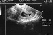
A normal placenta has a spongy structure, but sometimes, towards the end of pregnancy, some areas of the placenta can seem to petrify - these petrifications are called petrification or placental calcifications. Such hardened areas of the placenta are no longer able to fully perform their functions, but usually, even with multiple areas of petrification, the remaining fragments of the placenta cope with the functions assigned to them. If premature aging of the placenta or petrification in the placenta occurs, then the doctor will carefully monitor the state of the fetal cardiac activity so that oxygen deficiency can be ruled out in time. To prevent hypoxia, the drugs Actovegin or Hofitol can be prescribed. These drugs are safe for the fetus and can be used during pregnancy.
Diseases of the placenta
Like any other organs, the placenta can also have certain diseases - infection of the placenta, its infarctions can occur, blood clots can form in the vessels, and even tumor areas. This doesn’t happen often, but it’s still worth knowing about this state of affairs. Infectious damage to the placenta, or placentitis, can be caused by various microbes that penetrate the placenta in different ways. They can be brought by the bloodstream, can penetrate through the fallopian tubes from the appendage area, or penetrate upward from the vagina. Microbes can also penetrate from the uterus itself, if before pregnancy it was a source of inflammation. This condition is dangerous due to disruption of placental functions, and requires active treatment.
Infarctions and thromboses of the placenta result in the death of pieces of the placenta and disruption of the functions of gas exchange and nutrient transport in them. This leads to dysfunction of the placenta.
people, what does it mean - the structure of the placenta is homogeneous, thickened, but small in area S? 25 weeks and got the best answer
Answer from
Most likely, this is a feature of the development of this placenta. I wanted to write everything on my own, but I still googled it and called my mother (an obstetrician-gynecologist with more than 40 years of experience in the maternity hospital). How many of these placentas has she seen in 40 years of childbirth...))
She said so. There are different “cakes” of the placenta. There are large in area, but quite thin. There are thicker ones, but smaller in area. All this is a variant of the norm. Much depends on the place of its attachment - in the bottom, along the front wall, back, with transition to the sides, with or without presentation. The area of the placenta will also depend on this. If the placenta had to “rise” to normal, then it will be larger in area, but thinner. If it was initially located well, it will most likely be smaller in area, but thicker.
Such structural features do not affect its functions in any way; the vascular network develops in both the first and second variants equally correctly.
Poor-severe thickening (edema) of the placenta. Poorly calcifications in the placenta (petrificates), areas of infarction, disruption of uteroplacental blood flow, suspensions in the waters. Premature aging of the placenta is generally a controversial issue based on ultrasound (but this is not relevant, you don’t have that)
The rest is just features of the development of a particular placenta? , without deteriorating its functions. This does not affect the child, so it’s no big deal.
Answer from 2 answers[guru]
Hello! Here is a selection of topics with answers to your question: people, what does it mean that the structure of the placenta is homogeneous, thickened, but small in area S? 25 weeks
The most important task of using ultrasound diagnostics (ultrasound) during pregnancy is to study the structure and thickness of the placenta. The placenta is sometimes called the “baby place”, since it is it that nourishes the fetus and creates all the necessary conditions for its normal growth and development. Through it, maternal nutrition reaches the fetus. In addition, it serves as a protective barrier for the unborn baby, creating an obstacle to the penetration of infections, poisons, toxins and other harmful substances from a woman’s blood into the womb.
Norms and deviations
Up to 30 weeks (less often - up to 27), the placenta is characterized by a smooth, homogeneous structure without any inclusions. The appearance of hyperechoic inclusions in its tissue indicates a sufficient degree of maturity of the placenta.
These inclusions are called calcifications, and appear mainly at 30-32 weeks, immediately before birth. If this happens earlier, it is regarded as a pathological process called calcification.
Calcifications in the placenta that appear before 27-30 weeks are rarely regarded as an individual feature and a unique norm. Especially if the tissue structure is extremely heterogeneous, and single inclusions quickly “multiply”.
Essentially, calcification is regarded as premature aging. "children's place", which is not typical for a healthy pregnant woman. The maturity of the placenta is normal just before birth, when its natural removal from the body is just around the corner. If this happens ahead of schedule, the woman’s pregnancy is regarded as pathological, and the patient herself can be immediately hospitalized for further treatment and preservation.
Where do calcifications come from in the “young” placenta?
A heterogeneous placenta with calcifications detected late in pregnancy is not a cause for concern. However, if the formation of stones began earlier, namely before 27-30 weeks, the doctor should put the patient under careful observation.
As you know, the placenta is an organ with excellent blood supply. After all, it is he who carries fresh blood enriched with oxygen and nutrients to the developing fetus. If any pathological process occurs in the body of a pregnant woman, entailing a narrowing of blood vessels and capillaries, then the areas that were provided with nutritious blood may cease to function and simply begin to die.
It is at the site of damaged vessels that calcium salts are deposited, i.e., the formation of calcifications.
Since the death of areas of the placenta suppresses its permeability, the natural functions of this organ are also irreversibly disrupted, and the stages of normal fetal development become questionable.
The main provoking factors for the development of placental calcification are:
- Bad habits of the expectant mother (a special, “leading” place in their list should be given to active smoking);
- Urogenital infections (in particular, STIs and STDs);
- Other pathologies of infectious origin suffered during pregnancy;
- Chronic non-infectious diseases of internal organs in a pregnant woman;
- Severe degrees of gestosis in later stages;
- Severe anemia (anemia) in the mother;
- Systemic diseases (pathologies of the endocrine, cardiovascular, respiratory and urinary systems);
- Some pathologies of the uterus (fibroids, endometriosis, developmental abnormalities).
Single calcifications in the placental tissue do not manifest themselves in any way in everyday life and during pregnancy.
They are identified only during random or routine ultrasound examination. A placenta with multiple calcifications necessarily manifests itself with characteristic signs. First of all, a woman may notice changes in the movements of the fetus - they become either too sharp and active, or sharply weakened.
Since the baby’s well-being in the womb deteriorates sharply, the fetal heartbeat is disrupted, which can be detected during CTG (cardiotocography). The child exhibits tachycardia or bradycardia. The pregnant woman herself also begins to feel unwell. In some cases, women in this condition are diagnosed with late gestosis.
Finding a heterogeneous structure of the placenta with calcifications, the supervising obstetrician-gynecologist raises the question of medical preservation on an individual basis, depending on related factors and disorders.
Complications
You must understand that a disorder such as premature aging of the placenta can lead to a number of serious complications for you and the fetus:
As can be seen from the list, "side effects" calcification may well be fatal for you and your family.
Therefore, if you were previously diagnosed with such a diagnosis, but the supervising specialist did not deign to take any adequate measures in relation to it, and your health is systematically deteriorating, it makes sense to immediately seek qualified help from an outside physician.
Differential diagnosis
In order to take rational therapeutic measures, the doctor must accurately determine the cause that led to calcifications in the placenta at a period of 27-32 weeks and earlier.
To determine the exact cause of the problem, you will need the following diagnostic procedures:
Determining the cause of calcification is a critical part of its adequate therapy. Only in this case can a specific provoking factor be completely eliminated, which means that the woman will be reliably protected from the progression of the disorder and the development of obstetric pathologies.
Placenta provides a connection between the mother and the developing fetus. Many clinical problems are associated with its pathology, although this cannot always be subsequently confirmed by morphological examination. The term “placental insufficiency” has long been used to explain fetal growth and developmental restriction despite the absence of anatomical or morphological changes in the placenta.
With a wider implementation color Doppler mapping (CDC) to assess the disturbance of maternal and uteroplacental blood flow began to reveal the true meaning of the term “placental insufficiency”.
Although normal anatomy of the placenta and the options for its development, as well as various pathological changes, have now been studied quite well; an important aspect is the correct interpretation of their echographic manifestations, which can be detected during examination of this short-lived organ.
A main attention Our articles will focus on the correlation of ultrasound and morphological data in both normal and pathological conditions of the placenta.
Development of the placenta
On early stages fertilized egg surrounded by chorionic villi, which are visualized during transvaginal scanning in the form of a hyperechoic rim at 4-4.5 weeks (last menstruation). At 5 weeks, the villi located on the side opposite the implantation area regress, forming a smooth membrane with a small number of vessels, called the smooth chorion.
Remaining villi continue to proliferate and form the early placenta. At 9-10 weeks, ultrasound examination begins to clearly visualize the diffuse granular echostructure of the placenta. This ultrasound picture is a reflection from the branching structures of the villous tree, immersed in the lacunae of maternal blood. Such an echographic image of the placental tissue generally remains throughout pregnancy, against which calcium deposits gradually begin to be detected as the gestational age increases.
Formation of calcium deposits(petrificates) in the placenta during pregnancy is a physiological process. During the first six months they are microscopic in size, and mascroscopically visualized plaques form in the third trimester, usually after 33 weeks.
Initially petrifications deposited in the basal lamina and interlobular septa, but can also be found in the intervillous space and subchorially. Calcium deposits are easily visualized during ultrasound examination in the form of hyperechoic inclusions that do not create a significant “acoustic shadow” effect behind them.
With a pronounced process petrification interlobular septa in the structure of the placenta, hyper-echoic ring-shaped structures begin to be determined.
Petrification of the placenta was studied using histological, chemical, radiographic and echographic methods. It was found that the frequency of their detection, starting from 29 weeks, increases exponentially with increasing gestational age. After 33 weeks, a certain degree of petrification is observed in more than 50% of cases. In a fully mature placenta, the process of calcium deposition stops.
More often petrification registered in women with a small number of births in history, which may be due to the level of calcium in the blood plasma of the pregnant woman. Placental calcification is more common if delivery occurs at the end of summer or with premature birth, when the highest concentrations of calcium are observed in the maternal blood.
Currently time for convincing evidence It has not yet been established that the presence of petrificates in the placenta has any pathological or clinical significance.
Where is the placenta located and what does it look like?
During a normal pregnancy, the placenta is located in the area of the uterine body, developing most often in the mucous membrane of its posterior wall. The location of the placenta does not significantly affect the development of the fetus. The structure of the placenta is finally formed by the end of the first trimester, but its structure changes as the needs of the growing baby change. From 22 to 36 weeks of pregnancy, the placenta increases in mass, and by 36 weeks it reaches full functional maturity. A normal placenta at the end of pregnancy has a diameter of 15-18 cm and a thickness of 2 to 4 cm.
What does the placenta do?
Firstly, gas exchange occurs through the placenta: oxygen penetrates from the maternal blood to the fetus, and carbon dioxide is transported in the opposite direction.
Secondly, the fetus receives nutrients through the placenta and gets rid of its waste products.
Thirdly, the placenta has immune properties, that is, it allows the mother’s antibodies to pass through to the child, providing its immunological protection, and at the same time retains the cells of the mother’s immune system, which, having penetrated the fetus and recognizing it as a foreign object, could trigger fetal rejection reactions. (However, speaking about the protective function of the placenta, we must keep in mind that it practically does not protect the child from drugs, alcohol, nicotine, medications, viruses - they all easily penetrate through it).
Fourthly, the placenta plays the role of an endocrine gland and synthesizes hormones.
After childbirth (the placenta together with the membranes of the fetus - the afterbirth - is normally born within 15 minutes after the birth of the child), the placenta must be examined by the doctor who delivered the child. Firstly, it is very important to make sure that the placenta was born entirely (that is, there is no damage to its surface and there is no reason to believe that pieces of the placenta remained in the uterine cavity). Secondly, the state of the placenta can be used to judge the course of pregnancy (whether there was abruption, infectious processes, etc.).
Low attachment of the placenta
Low placental attachment is a fairly common pathology: 15-20%. If a low location of the placenta is determined after 28 weeks of pregnancy, they speak of placenta previa, since in this case the placenta at least partially covers the uterine os. However, fortunately, only 5% have a low-lying placenta until 32 weeks, and only a third of these 5% have a low-lying placenta by 37 weeks.
Placenta previa
If the placenta reaches the internal os or overlaps it, they speak of placenta previa (that is, the placenta is located in front of the presenting part of the fetus). Placenta previa most often occurs in repeatedly pregnant women, especially after previous abortions and postpartum diseases. In addition, placenta previa is promoted by tumors and abnormal development of the uterus, and low implantation of the fertilized egg. Ultrasound detection of placenta previa in the early stages of pregnancy may not be confirmed in later stages. However, such an arrangement of the placenta can provoke bleeding and even premature birth, and therefore is considered one of the most serious types of obstetric pathology.
Placenta accreta
During the formation of the placenta, chorionic villi “invade” the mucous membrane of the uterus (endometrium). This is the same membrane that is rejected during menstrual bleeding - without any damage to the uterus and to the body as a whole. However, there are cases when villi grow into the muscle layer, and sometimes throughout the entire thickness of the uterine wall. Placenta accreta is also facilitated by its low location, because in the lower segment of the uterus the chorionic villi “deepen” into the muscle layer much more easily than in the upper sections.
Tight attachment of the placenta
In fact, dense placenta attachment differs from placenta accreta in the shallower depth of chorionic villi growth into the uterine wall. Just like placenta accreta, tight attachment often accompanies placenta previa or low-lying placenta. Unfortunately, it is possible to recognize placenta accreta and tight attachment (and distinguish them from each other) only during childbirth. If the placenta is firmly attached and accreta occurs in the afterbirth period, the placenta does not separate spontaneously. When the placenta is tightly attached, bleeding develops (due to detachment of areas of the placenta); There is no bleeding with placenta accreta. As a result of accreta or tight attachment, the placenta cannot separate in the third stage of labor. In the case of a tight attachment, they resort to manual separation of the placenta - the doctor delivering the baby inserts his hand into the uterine cavity and separates the placenta.
Placental abruption
As noted above, placental abruption can accompany the first stage of labor with a low-lying placenta or occur during pregnancy with placenta previa. In addition, there are cases when premature detachment of a normally located placenta occurs. This is a severe obstetric pathology, observed in 1-3 out of a thousand pregnant women.
Manifestations of placental abruption depend on the area of detachment, the presence, size and speed of bleeding, and the woman’s body’s reaction to blood loss. Small detachments may not manifest themselves in any way and may be detected after childbirth when examining the placenta.
If the placental abruption is insignificant, its symptoms are mild, if the amniotic sac is intact, it is opened during childbirth, which slows down or stops the placental abruption. A pronounced clinical picture and increasing symptoms of internal bleeding are indications for a cesarean section (in rare cases, it is even necessary to resort to removal of the uterus - if it is soaked in blood and does not respond to attempts to stimulate its contraction).
If, during placental abruption, childbirth occurs through the natural birth canal, then a manual examination of the uterus is mandatory.
Early maturation of the placenta
The placenta goes through four stages: formation, growth, maturity, aging. Each stage corresponds to a certain degree of maturity:
Formation - 0
Maturity - 2
Aging - 3
There is premature maturation of the placenta - a condition when the placenta reaches the first or second degree of maturity ahead of time. This condition in itself is not dangerous, but if it is detected, the placenta must be closely monitored, since during such a pregnancy premature aging of the placenta is possible.
Premature aging is said to occur when the placenta reaches the third degree of maturity before 37 weeks of pregnancy. With premature aging of the placenta, fetoplacental insufficiency may occur, so it is necessary to do a CTG. However, if you have discovered aging of the placenta, then you should not worry too much, since the compensatory possibilities are quite large. Therefore, as a rule, premature aging of the placenta does not affect the condition of the fetus in any way, and the child is born at term.
Depending on the pathology of pregnancy, insufficiency of placental function when it matures too early is manifested by a decrease or increase in the thickness of the placenta. Thus, a “thin” placenta (less than 20 mm in the third trimester of pregnancy) is characteristic of late toxicosis, threat of miscarriage, fetal malnutrition, while in case of hemolytic disease and diabetes mellitus, a “thick” placenta (50 mm or more) indicates placental insufficiency. . Thinning or thickening of the placenta indicates the need for therapeutic measures and requires repeated ultrasound examination.
Late maturation of the placenta
It is observed rarely, more often in pregnant women with diabetes mellitus, Rh conflict, as well as in congenital malformations of the fetus. Delayed placental maturation leads to the fact that the placenta, again, inadequately performs its functions. Often, late maturation of the placenta leads to stillbirth and mental retardation in the fetus. Reduction in the size of the placenta. There are two groups of reasons leading to a decrease in the size of the placenta. Firstly, it may be a consequence of genetic disorders, which is often combined with fetal malformations (for example, Down syndrome). Secondly, the placenta may “fall short” in size due to the influence of various unfavorable factors (severe gestosis in the second half of pregnancy, arterial hypertension, atherosclerosis), ultimately leading to a decrease in blood flow in the vessels of the placenta and to its premature maturation and aging (see. higher). In both cases, the “small” placenta cannot cope with its responsibilities of supplying the baby with oxygen and nutrients and ridding him of metabolic products.
Increased placenta size
Placental hyperplasia occurs with Rh conflict, severe anemia in a pregnant woman, diabetes mellitus in a pregnant woman, syphilis and other infectious lesions of the placenta during pregnancy (for example, with toxoplasmosis), etc. There is little point in listing all the reasons for an increase in the size of the placenta, but it must be borne in mind that when this condition is detected, it is very important to establish the cause, since it is this that determines the treatment. Therefore, you should not neglect the studies prescribed by your doctor - after all, the consequence of placental hyperplasia is the same placental insufficiency, leading to delayed intrauterine development of the fetus.
Placental hypoplasia
Hypoplasia is a condition when the placenta is significantly smaller than normal for a given period. We are talking about a significant reduction, because there are individual characteristics. Reduction of the placenta can be primary or secondary. Primary reduction is most often caused by various genetic abnormalities, in which case the fetus itself often has genetic diseases. Primary hypoplasia is a fairly rare occurrence. The most common type is secondary placental hypoplasia. It can be caused by stress, smoking, poor nutrition of the mother, or an infectious disease suffered during pregnancy. In addition, secondary hypoplasia can cause gestosis. With a reduced placenta, the supply of nutrients and oxygen to the fetus may decrease.
Membranous placenta
This is a very thin but extensive placenta. Its diameter can reach 40 cm. Most often, this pathology occurs due to a chronic inflammatory process in the uterus. With this pathology, fetoplacental insufficiency (FPI) may occur.
Petrification of the placenta
Normally, the placenta has a soft, spongy structure. Sometimes, at the end of pregnancy, some parts of the placenta “petrify.” These pebbles are called petrificates or calcifications. Hardened areas of the placenta are not able to perform their functions, but, as a rule, even with multiple petrifications, the remaining part of the placenta copes with its work normally.
In case of premature aging and/or petrification of the placenta, the doctor will closely monitor the cardiac activity of the fetus to exclude oxygen starvation (this occurs quite rarely). To prevent oxygen starvation, hofitol or Actovegin can be prescribed. These drugs do not affect the fetus and are therefore absolutely harmless during pregnancy.


